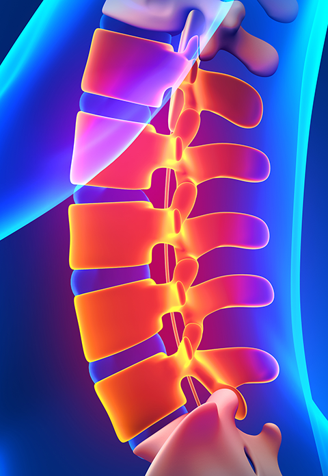
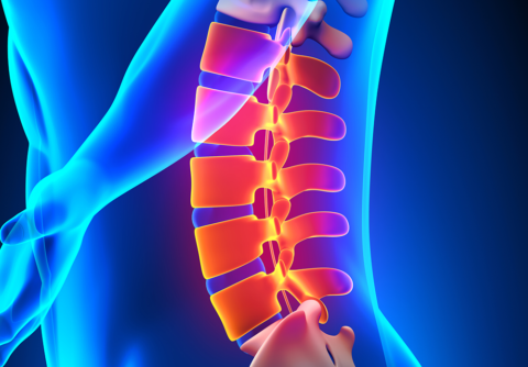
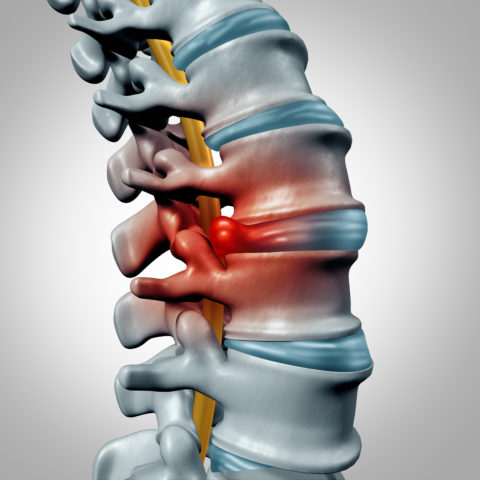
The facet joints, also referred to as zygapophyseal joints or z-joints, is a true joint in the posterior (closer to the back) elements of the spine formed by the articulation between all the spinal vertebral levels. Each level has two facet joins, one on both sides. The Facet joint contains two smooth surfaces covered in cartilage, and the joint contains joint fluid. There is a joint capsule around the joint to hold the joint fluid in place. The facet joints allow for the majority of flexibility of the spine.
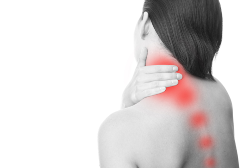
In general, most medical studies that investigate facet joint disease state that it accounts for 15-40% of low back pain and 40-60% of axial neck pain. If the facet joints are damaged for any reason pain may develop. Injury may result from trauma, arthritis, repetitive injury or other causes. Individuals with facet joint disease usually present with pain over the affected joint, and pain may refer to different area. There may be associated crepitus (grinding, popping and clicking sounds) with movement of the area. Pain is usually worse with movement of the affected area, and worse when extending and rotating the affected segment. The facet joints in the neck (cervical facet joints) may refer pain into the head, which causes headaches. There have been been very eloquent studies completed which maps the referral pattern of the cervical facets, and they can refer to the head, neck or shoulder blade and upper arm area. Facet joints in the mid back (thoracic area) usually cause pain over the affected segment. Lumbar (low back) facet joints can cause pain over the joint, but also refer pain into the buttock and down into the thigh.
Imaging studies can be helpful, such as x-ray, CT scan and MRI, as they may show abnormalities in the facet joint, but in some cases there may be “normal” findings on imaging and the imaging does not always correlate with the extend of pain. Diagnostic facet joint blocks are still the gold standard for diagnosing pain from facet joint disease.

